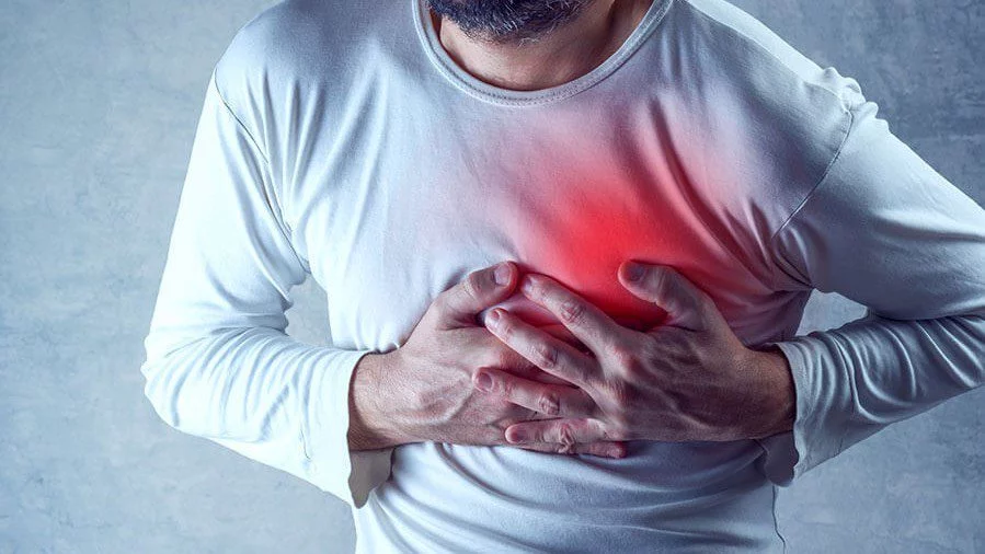
The setting in which chest pain occurs provides one of the most important clue for the evaluation. The history of chest pain is more important than the actual physical examination. Important features of history of chest pain usually involves following main points:
- Duration
- Location
- Quality
- Radiation
- Frequency
- Aggravating and relieving factors.
There are few important aspects that physician and patient should remember so as to take quick action and come to quick clinical
Pain described as tightness or heaviness, upper abdominal pain, nausea vomiting, bradycardia (decreased rate of heartbeat), fall in blood pressure (dizziness, fainting) can be due to Angina, acute coronary syndrome or inferoposterior wall ischemia.
If the patient can pin point the exact location of the pain which is sharp and knife like associated or reproduced by changes in position or by touching it is less likely that the pain has cardiovascular origin.
Nitroglycerin (dilates the smooth muscles) can worsen the pain (Gastroesophageal reflux disease) or it may decrease the pain within minutes (Transient ischemia, Esophageal spasm).
Physical Examination: Read Causes of Chest Pain
Initial Impression: Diaphoresis, Increased rate of breathing or an anxious patient may give clue about life threatening underlying cause of chest pain.
Blood Pressure: Difference of 20 mm of Hg or more in both the arms maybe suggestive of Aortic dissection.
Hypo-tension: May be due to massive pulmonary embolism or cardiac shock.
Increased rate of breathing (Tachypnea) and Increase heart beat (Tachycardia) is very non specific but is always present in pulmonary embolism. Fever may suggest pneumonia. Esophageal rupture also presents with chest pain and mediastinitis.
If there is pain while palpating (touching the chest wall) the chest pain could of musculoskeletal in origin.
New murmurs and abnormal heart sounds:
RBBB (right bundle branch block) & right ventricular infarction: Wide physiological splitting of second heart sound with inspiration.
LBBB (left bundle branch block), anterior or lateral wall infarction: New paradoxical splitting of second heart sound.
Fourth heart sound: can occur with angina or infarction.
Significant Aortic regurgitation is almost always associated with aortic dissection. Mitral regurgitation can occur in patients with angina or infarction with papillary muscle dysfunction.
Lung Auscultation for Crackles, asymmetrical (spontaneous pneumothorax) or absent breath sounds (pneumothorax and pleural effusion).
Extremities should be examined for swelling (unilateral)- pulmonary embolism or for pulses (absence pf pedal pulse –Aortic dissection)
Tests: Investigations For Evaluating Chest Pain
Read Tests for Chest Pain in detail
ECG (Electrocardiograph) is the single most initial test to rule out cardiac cause of chest pain and should be done with 12 leads immediately after initial assessment and recording of vitals. Patients presenting with acute chest pain and normal ECG the chances of chest pain being cardiac in origin is about 10 percent and in some studies 1-2 percent. So it is very important to record ECG so as to differentiate between cardiac and non cardiac causes of chest pain.
Cardiac Enzymes: CK (MB), cardiac troponins play a vital role in workup of the patient to determine whether the chest pain is cardiac in origin, angina or ischemia. They are also important markers of recent myocardial infarction.
Chest x ray: Can tell the origin of the chest pain, if it is due to some lung infection, aortic dissection, esophageal rupture, pulmonary embolism.


